Home

tassa di iscrizione revisione emorragia mouse hippocampus coronal section spirito Minatore ascesso

BioRender | Life Science Icons

Schema of a coronal (a) section of the mouse brain showing the... |  Download Scientific Diagram
Schema of a coronal (a) section of the mouse brain showing the... | Download Scientific Diagram

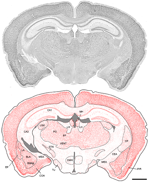
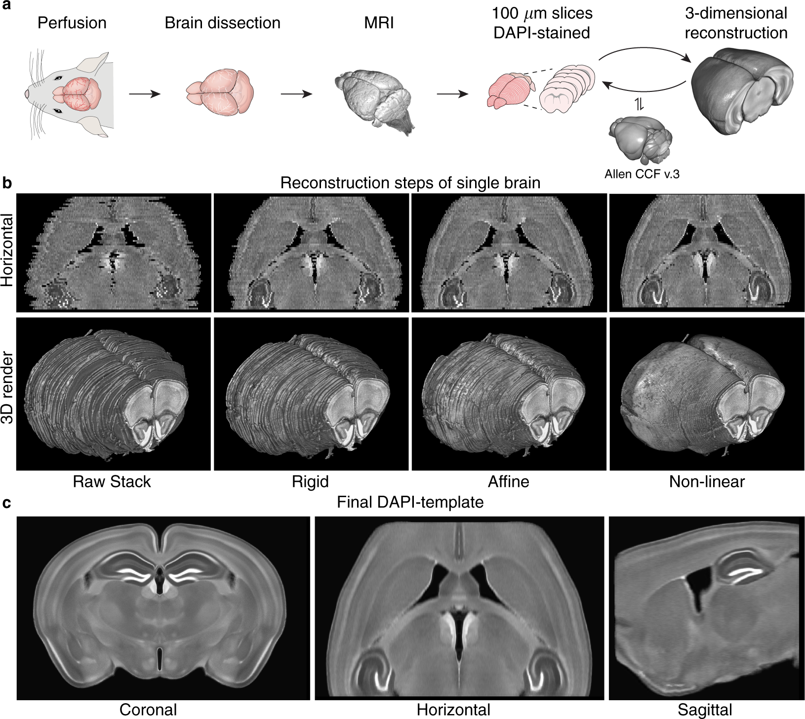
A three-dimensional, population-based average of the C57BL/6 mouse brain  from DAPI-stained coronal slices | Scientific Data
A three-dimensional, population-based average of the C57BL/6 mouse brain from DAPI-stained coronal slices | Scientific Data
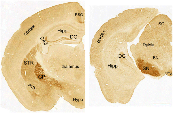
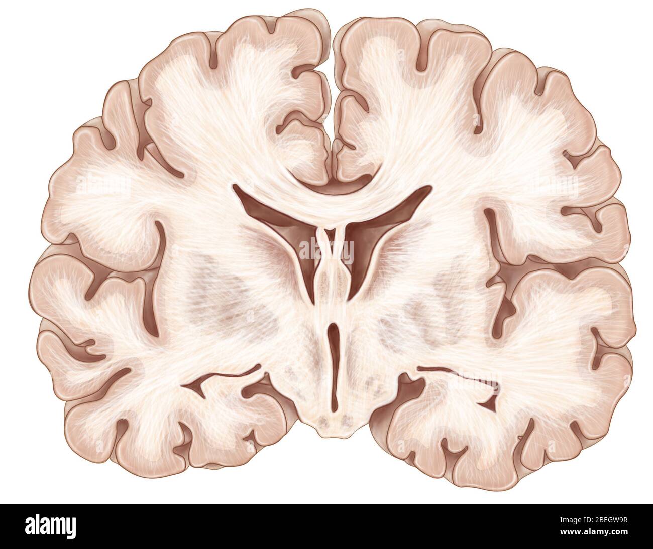
Brain, Coronal Section Stock Photo - Alamy

FMRP in Mouse brain | College of Medicine

Figure 2 | Mutant Huntingtin Causes a Selective Decrease in the Expression  of Synaptic Vesicle Protein 2C | SpringerLink
Figure 2 | Mutant Huntingtin Causes a Selective Decrease in the Expression of Synaptic Vesicle Protein 2C | SpringerLink

Figure 1. | The Adult Mouse Hippocampal Progenitor Is Neurogenic But Not a  Stem Cell | Journal of Neuroscience
Figure 1. | The Adult Mouse Hippocampal Progenitor Is Neurogenic But Not a Stem Cell | Journal of Neuroscience

The Mouse Brain Library

Mouse Brain Atlases
Mouse Brain Atlases

Mice transgenically overexpressing sulfonylurea receptor 1 in forebrain  resist seizure induction and excitotoxic neuron death | PNAS
Mice transgenically overexpressing sulfonylurea receptor 1 in forebrain resist seizure induction and excitotoxic neuron death | PNAS

Geometry of the coronal and sagittal sections of a mouse brain used... |  Download Scientific Diagram
Geometry of the coronal and sagittal sections of a mouse brain used... | Download Scientific Diagram

Mouse C57 Brain Coronal Paraffin Tissue Sections – Zyagen

A coronal mouse brain section showing probe placements (illustrated by  vertical lines) in the nucleus of mice used in the present study.
A coronal mouse brain section showing probe placements (illustrated by vertical lines) in the nucleus of mice used in the present study.

Molecular atlas of the adult mouse brain

Coronal section of the mouse brain illustrating the main areas of the... |  Download Scientific Diagram
Coronal section of the mouse brain illustrating the main areas of the... | Download Scientific Diagram

High Resolution Mouse Brain Atlas - Methods

Serial coronal sections of E18.5 mouse brain. Wild type... | Download  Scientific Diagram
Serial coronal sections of E18.5 mouse brain. Wild type... | Download Scientific Diagram

BioRender | Life Science Icons

PDF] 3-D Mouse Brain Model Reconstruction from a Sequence of 2-D Slices in  Application to Allen Brain Atlas | Semantic Scholar
PDF] 3-D Mouse Brain Model Reconstruction from a Sequence of 2-D Slices in Application to Allen Brain Atlas | Semantic Scholar
Automatic navigation system for the mouse brain | bioRxiv

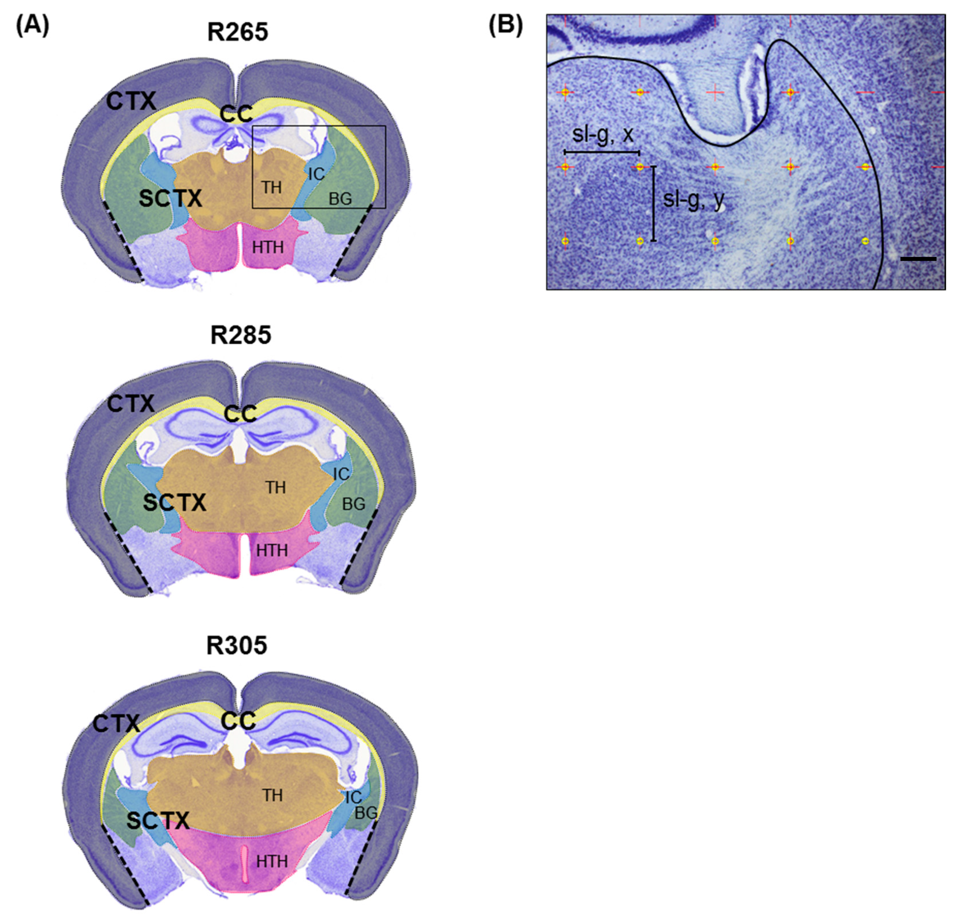
Cells | Free Full-Text | Stereological Investigation of Regional Brain  Volumes after Acute and Chronic Cuprizone-Induced Demyelination | HTML
Cells | Free Full-Text | Stereological Investigation of Regional Brain Volumes after Acute and Chronic Cuprizone-Induced Demyelination | HTML

View Image
View Image

Pin on PhD
Pin on PhD